![]() Figure 4 of
Chen, Mol Vis 2007;
13:374-387.
Figure 4 of
Chen, Mol Vis 2007;
13:374-387.
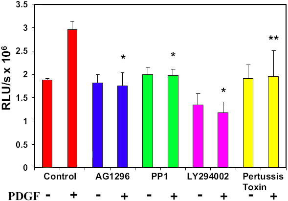
Figure 4. Determination of PDGF-stimulated cell proliferation in the presence or absence of inhibitors by BrdU incorporation assay
DNA synthesis induced by PDGF (1 ng/ml, 60 min) in cells with and without pretreatment of PDGF receptor inhibitor, AG1296 (20 μM, overnight), Src inhibitor, PP1 (10 μM, 30 min), PI3K inhibitor, LY294002 (30 μM, 30 min) or GPCR inhibitor, pertussis toxin (500 ng/ml, overnight) was measured by BrdU incorporation. Control cells (no inhibitor), were included for comparison. The data are expressed as relative luminescence unit (RLU)/sec, with mean±SD (n=12). The results are from 3 separate experiments. Each p value was obtained by using inhibitor (with PDGF) against control (with PDGF). The asterisk indicates a p<0.001 and the double asterisk denotes a p<0.026.