![]() Figure 2 of
Chen, Mol Vis 2007;
13:374-387.
Figure 2 of
Chen, Mol Vis 2007;
13:374-387.
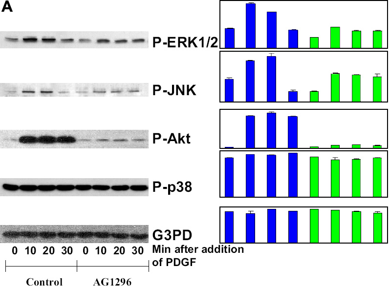
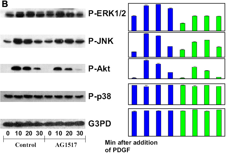
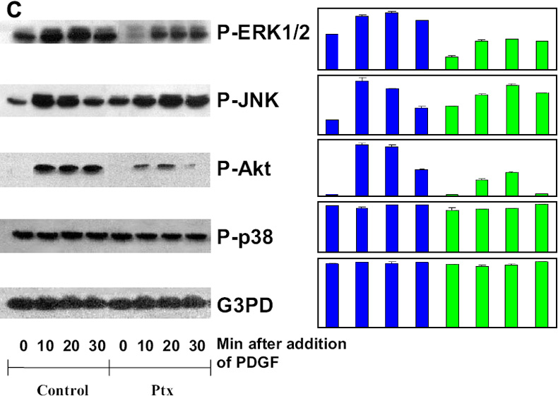
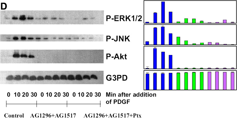
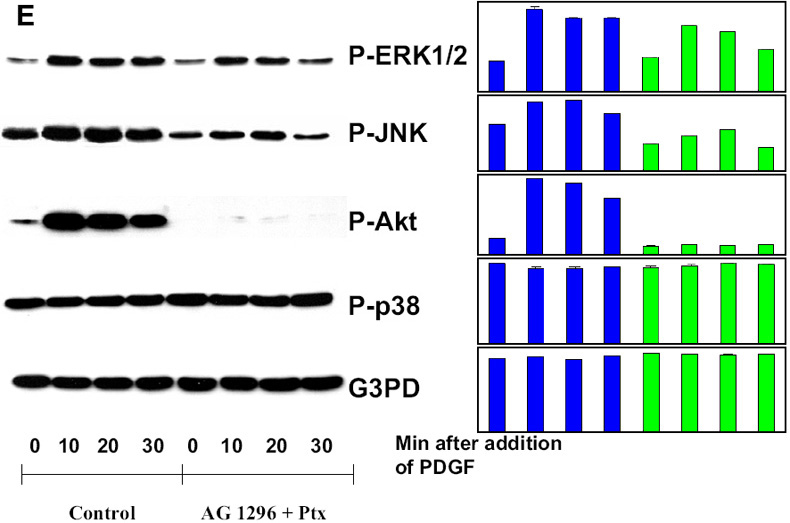
Figure 2. Effect of receptor inhibition on PDGF-activated MAP kinases in HLE B3 cells
Serum-deprived cells with and without inhibitor preloading were stimulated with PDGF (1 ng/ml) for 0, 10, 20, and 30 min, harvested and then lysed in lysis buffer. Cell lysates were applied on 10% SDS-PAGE, transblotted and probed with specific antibodies to P-ERK1/2, P-JNK, P-Akt, P-p38, and G3PD, respectively. A: Cells were stimulated with PDGF with and without preloading (2.5 h) of PDGF receptor inhibitor, AG1296 (20 μM). B: Cells were stimulated with PDGF with and without preloading (2.5 h) with EGF receptor inhibitor, AG1517 (1 μM). C: Cells were stimulated with PDGF with and without preloading (2.5 h) with pertussis toxin (Ptx, 500 ng/ml). D: Cells were stimulated with and without preloading (2.5 h) with AG1296 (20 μM) + AG1517 (1 μM), or AG1296 (20 μM) + AG1517 (1 μM) + Ptx (500 ng/ml). E: Cells were stimulated with and without preloading (overnight) with AG1296 (20 μM) + Ptx (250 ng/ml). Cells without treatment of inhibitors but with stimulation of PDGF (1 ng/ml) were used as the controls. The bar graph with averaged pixel values of the band intensities for each western blot is shown. Data presented are a typical representation of triplicate experiments.