![]() Figure 4 of
Biswas, Mol Vis 2007;
13:345-359.
Figure 4 of
Biswas, Mol Vis 2007;
13:345-359.
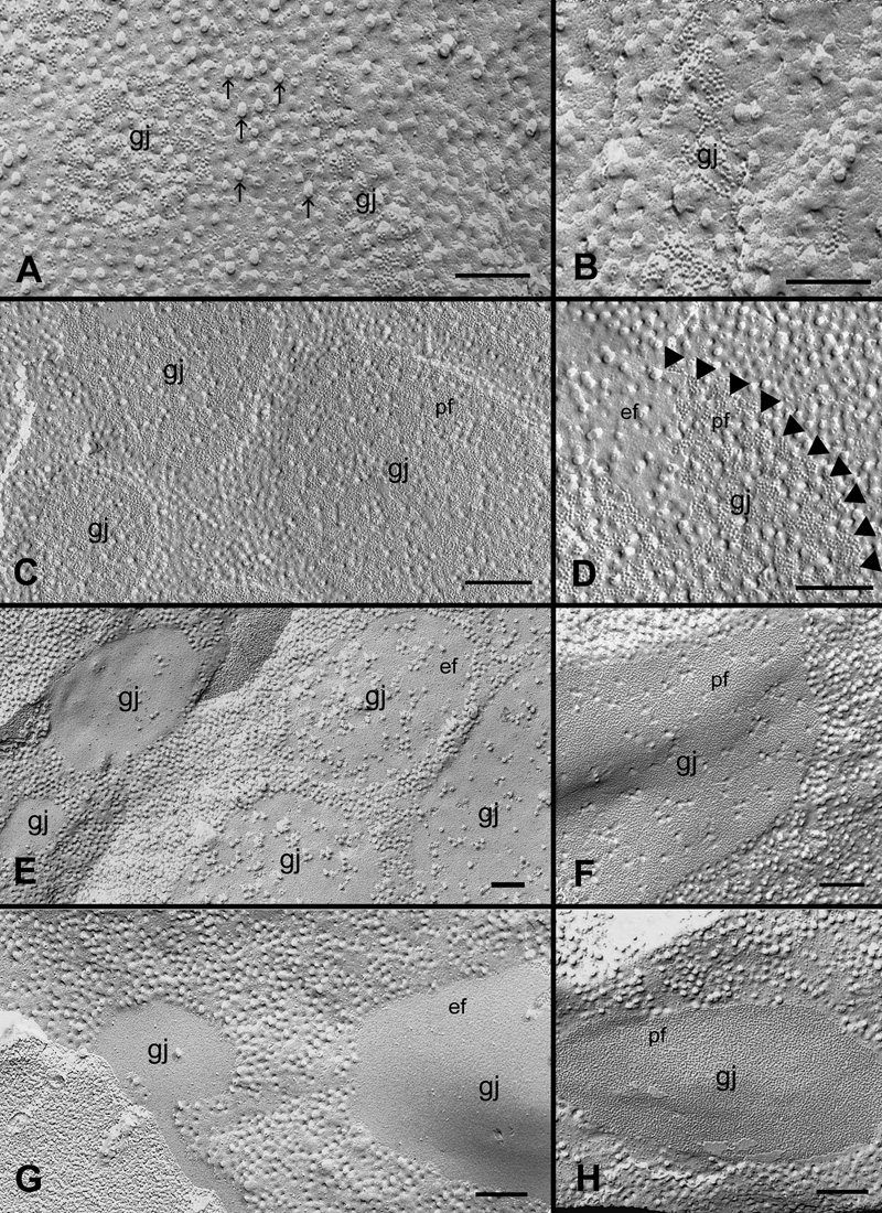
Figure 4. Heterogeneous distribution of cholesterol in gap junction plaques in the outer cortical fiber cells
Gap junctions in the outer cortex (0-200 μm deep) contain different amounts of cholesterol as determined by filipin cytochemistry and freeze-fracture TEM. Newly formed gap junctions (A and B) and well-formed cholesterol-rich gap junctions (C and D) contain a large number of filipin-cholesterol complexes (FCCs). Some FCCs (25-35 nm particles) in the P-face of the non-junctional membrane are indicated by the arrows in A. At high magnification, the border of such cholesterol-rich gap junction is outlined by arrowheads for a clearer visualization (D). Cholesterol-intermediate gap junctions contain considerably less FCCs (E and F). Cholesterol-poor or -free gap junctions contain only a few or no FCCs in the junctional plaques (G and H). pf, P-face of the membrane; ef, E-face of the membrane. The scale bars are equal to 200 nm.