![]() Figure 2 of
Biswas, Mol Vis 2007;
13:345-359.
Figure 2 of
Biswas, Mol Vis 2007;
13:345-359.
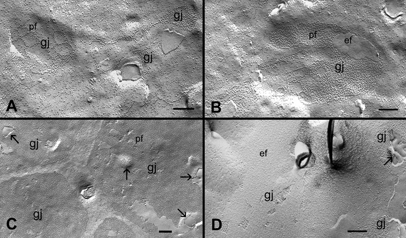
Figure 2. Different structural configurations of gap junctions in various cortical regions of the embryonic chicken lens
Representative micrographs show different structural configurations of gap junctions (gj) found in various cortical regions in the embryonic chicken lens. A and B: The assembly of forming gap junction plaques in the superficial fibers. C: A cluster of well-formed gap junction plaques in the outer cortical fibers. D: Several mature gap junction plaques found in the inner cortical fibers. Note the presence of vesicular structures (arrows) associated with gap junctions found in both outer and inner cortical fibers. pf, P-face of the membrane; ef, E-face of the membrane. The scale bars are equal to 200 nm.