![]() Figure 1 of
Biswas, Mol Vis 2007;
13:345-359.
Figure 1 of
Biswas, Mol Vis 2007;
13:345-359.
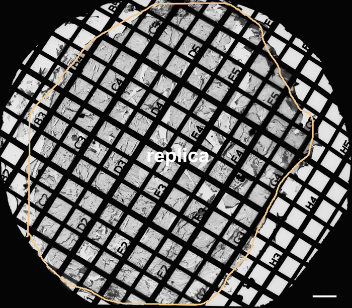
Figure 1. A large intact replica on the Girder finder grid with index number for systematic examinations of fiber gap junctions in the embryonic chicken lens
A low magnification shows an overview of a large intact replica (outlined by circle line) on the Girder finder grid with index number for systematic examinations of fiber gap junctions from outer to inner cortical regions of the embryonic chicken lens at E15. The peripheral area along the circle line represents the superficial surface, and the center represents the nucleus of the lens. This freeze-fracture replica was prepared from a 300 μm lens slice initially cut with a Vibratome for the longitudinal orientation of cortical fiber cells. In this study, initial identifications of various lens regions were made and recorded using the index number (the distance between two parallel grid bars is 100 μm), followed by thorough examinations of the structures of interest in each region. The scale bar is equal to 100 μm.