![]() Figure 4 of
Thomas, Mol Vis 2007;
13:337-344.
Figure 4 of
Thomas, Mol Vis 2007;
13:337-344.
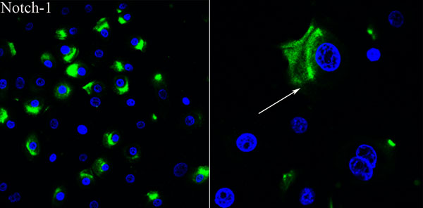
Figure 4. Characterization of epithelial outgrowth from limbal explants
Notch-1 showed expression in the cytoplasm while the signal disappeared in dividing cells. The arrow points to a cell with positive staining. In the panels, the green fluorescence is FITC conjugated anti mouse/rat IgG, and the blue fluorescence is nuclei counterstained with DAPI. The right panel is an enlarged view of the left panel.