![]() Figure 3 of
Thomas, Mol Vis 2007;
13:337-344.
Figure 3 of
Thomas, Mol Vis 2007;
13:337-344.
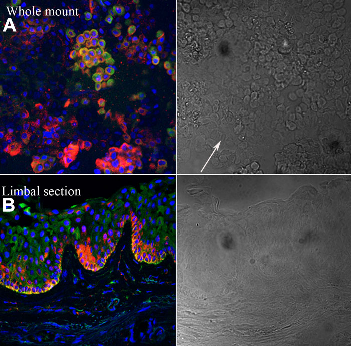
Figure 3. Colocalization of ABCG2 and Notch-1 in the human Limbo-corneal epithelium
A: When the whole mount was double stained with ABCG2 and Notch-1, subsets of cells were positive for both. Note that most Notch-1 positive cells (green fluorescence: FITC conjugated anti rat IgG) coexpressed ABCG2 (red fluorescence: rhodamine red X conjugated anti mouse IgG and nuclear counter stain with DAPI). B: When frozen sections were double stained they showed colocalization of ABCG2 and Notch-1 in a subset of cells in the limbal basal region.The right panels show representative phase contrast micrographs for A and B.