![]() Figure 2 of
Thomas, Mol Vis 2007;
13:337-344.
Figure 2 of
Thomas, Mol Vis 2007;
13:337-344.
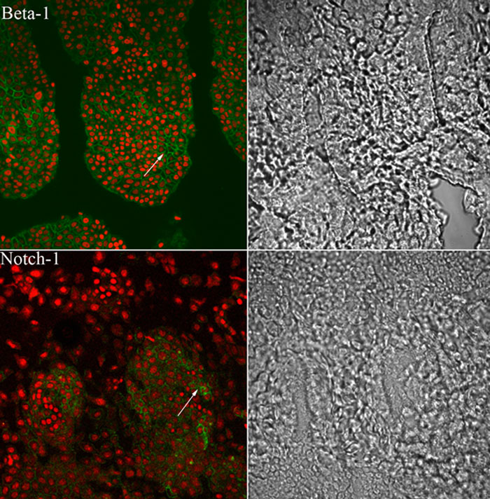
Figure 2. Localization of integrin β1 and Notch-1 in the whole mount
The immunoflurescence staining showing the limbal area with the palisade of Vogt environment in the left panel. Immunostaning for integrin β1 showed a group of bright cells in the limbal area (see arrow). Notch-1 expression was prominent only in the putative stem cells in the palisades of Vogt. (In the panels, green fluorescence represents FITC conjugated anti mouse/rat IgG and the red fluorescence shows nuclear counter stain with propidium iodide). The phase contrast micrographs in the right panels show the limbal architecture.