![]() Figure 7 of
Warburton, Mol Vis 2007;
13:318-329.
Figure 7 of
Warburton, Mol Vis 2007;
13:318-329.
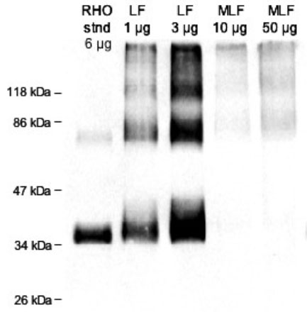
Figure 7. Rhodopsin immunoblot
Immunoblot following SDS-PAGE of 1 and 3 μg of total lipofuscin (LF) protein and 10 and 50 μg of total melanolipofuscin (MLF) protein. Shown for comparison is 6 μg of protein from photoreceptor cell membranes enriched from human retina. Rhodopsin runs on SDS-PAGE as a mixture of the monomer (about 30 kDa), dimer (about 60 kDa), and trimer (about 90 kDa). Although rhodopsin is seen to be present in abundance in LF granules, no significant quantity of rhodopsin is detected in MLF granules.