![]() Figure 6 of
Warburton, Mol Vis 2007;
13:318-329.
Figure 6 of
Warburton, Mol Vis 2007;
13:318-329.
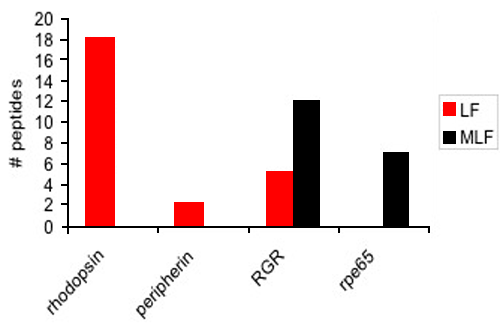
Figure 6. Semiquantitative analysis of photoreceptor- and retinal pigment epithelium-specific proteins in lipofuscin and melanolipofuscin granules
Spectral counting was performed on two photoreceptor-specific proteins, rhodopsin and peripherin, and two retinal pigment epithelium (RPE)-specific proteins, RGR and rpe65, in 4 gel slices from lipofuscin (LF) and melanolipofuscin (MLF) 1D gels. Photoreceptor-specific proteins were only identified in LF granules, while RPE-specific proteins were mainly identified in MLF granules. Although RGR was identified in LF granules it appeared to be about 58% less abundant than in MLF granules. This supports the hypothesis that LF granules originate from photoreceptors while MLF granules appear to originate from autophagy of RPE cells.
