![]() Figure 4 of
Warburton, Mol Vis 2007;
13:318-329.
Figure 4 of
Warburton, Mol Vis 2007;
13:318-329.
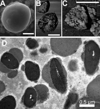
Figure 4. Microscopic structure of melanolipofuscin
A: Scanning electron micrograph of melanolipofuscin (MLF), showing nearly spherical granules with some surface features. B, C: Atomic force micrographs (phase images) showing MLF granules to be aggregates of about 200 nm and about 50 nm substructures. D: Transmission electron micrograph of MLF, shows these granules to contain inclusions of higher density, demonstrating that these granules are complexes of lipofuscin and melanin. Each bar represents 0.5 μm.