![]() Figure 2 of
Liu, Mol Vis 2007;
13:309-317.
Figure 2 of
Liu, Mol Vis 2007;
13:309-317.
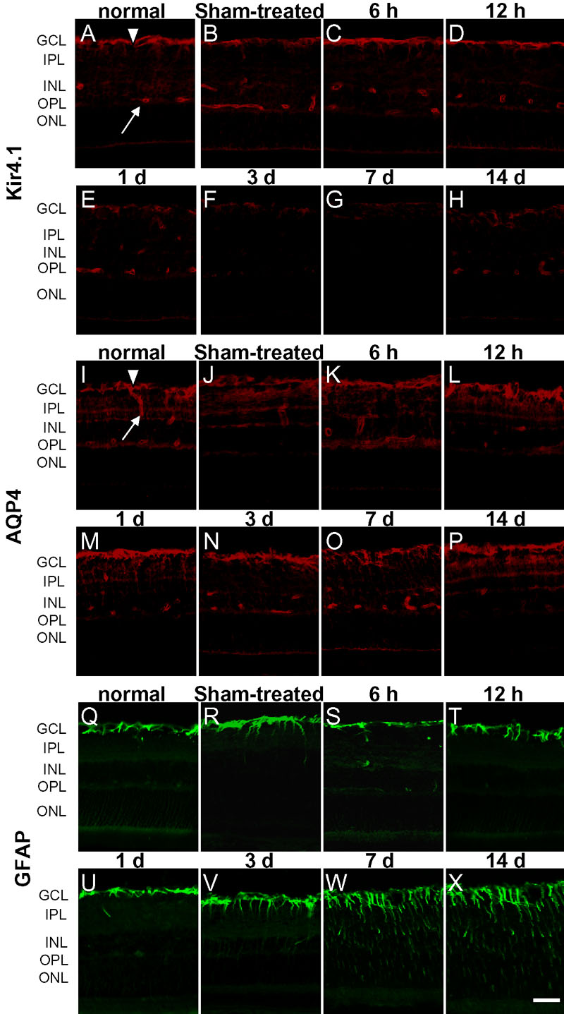
Figure 2. Immunohistochemical detection of Kir4.1, aquaporin-4, and anti-glial fibrillary acidic protein in the retina
In the normal eyes, Kir4.1 (A) and aquaporin-4 (AQP4; I) were enriched in the endfoot membranes facing the vitreous body (arrowheads) and retinal blood vessels (arrows). Staining for AQP4 (K-P) maintained the same pattern during the different stages of endotoxin-induced uveitis (EIU), and only a slight reduction in immunostaining was seen in the inner plexiform at 1-7 day after lipopolysaccharide (LPS) injection. Kir4.1 (C-H) immunoreactivity decreased significantly from one day after LPS injection, had almost disappeared at 3-7 day after injection, and had partially recovered by 14 days. Anti-glial fibrillary acidic protein (GFAP; Q-X) was predominantly found in astrocytes in the retinas of the untreated controls. Seven days and 14 days after intravitreal LPS injection, GFAP (W-X) immunoreactivity was significantly increased in Müller cells. In the retinas of sham-treated eyes, the immunostaining for Kir4.1 (A-H) was unchanged at 3 day after phosphate-buffered saline (PBS) treatment (B). AQP4 immunoreactivity was unchanged at one day after PBS treatment (J). GFAP immunoreactivity was mildly increased at 7 day after PBS treatment (R). GCL indicates ganglion cell layer; INL indicates inner nuclear layer; IPL indicates inner plexiform layer; ONL indicates outer nuclear layer; OPL indicates outer plexiform layer. Scale bar represents 20 μm.