![]() Figure 1 of
Liu, Mol Vis 2007;
13:309-317.
Figure 1 of
Liu, Mol Vis 2007;
13:309-317.
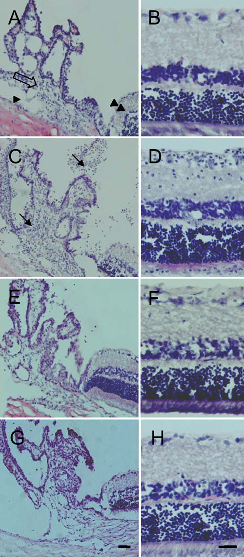
Figure 1. Hematoxylin-eosin stained eyes
A, B: No inflammatory cells were seen in the anterior chamber (arrowhead), iris-ciliary body (blank arrow), posterior chamber and retina (double arrowhead) of untreated control eyes. C, D: Inflammatory cells (arrows) were seen in the anterior chamber, iris-ciliary body, posterior chamber and retina one day after lipopolysaccharide (LPS) injection. E, F: Inflammation had almost subsided and only a few inflammatory cells remained seven days after LPS injection. G, H: No inflammatory cells were seen in the sections one day after phosphate-buffered saline treatment. Scale bar represents 20 μm.