![]() Figure 8 of
Khalyfa, Mol Vis 2007;
13:293-308.
Figure 8 of
Khalyfa, Mol Vis 2007;
13:293-308.
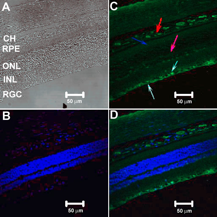
Figure 8. Confocal immunocytochemical localization of complement factor H complement factor H in the adult rat retina
A DIC image of retina showing various layers of rat retina (A). The nuclear staining by Hoechst stain is shown in (B). Intense factor H immunolabeling (C) was detected in retinal ganglion cell (RGC) layer (white arrow) including the nerve fiber layer (fluorescent blue arrow). Factor H labeling was also detected in the inner plexiform layer (purple arrow), photoreceptors (blue arrow), and the choroids (red arrow). The merged image of B and C is shown in D. INL indicates inner nuclear layer; ONL indicates outer nuclear layer; RPE indicates retinal pigment epithelium layer; and CH indicates choroids.