![]() Figure 5 of
Khalyfa, Mol Vis 2007;
13:293-308.
Figure 5 of
Khalyfa, Mol Vis 2007;
13:293-308.
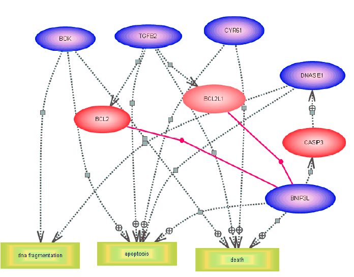
Figure 5. Biological pathway for the differentially expressed neuronal cell death genes in serum-deprived retinal ganglion cells
Pathways were identified by incorporating the microarray results (genes which are differentially expressed at 24, 48, and 96 h) into the Pathway Assit software. The pathway was constructed on this software by searching for the shortest path to connect the genes of interest by other genes or cell processes with which they interacted through expression or regulation only. Three major biologic processes are identified (apoptosis, death, and DNA fragmentation) and are represented by yellow rectangles. Blue ovals denote genes identified as neuronal cell death, and red ovals new genes connected to this pathway.