![]() Figure 1 of
Khalyfa, Mol Vis 2007;
13:293-308.
Figure 1 of
Khalyfa, Mol Vis 2007;
13:293-308.
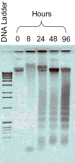
Figure 1. Apoptotic DNA fragmentation in serum-deprived retinal ganglion cell-5 samples
DNA was extracted and electrophoresed in parallel on the same agarose gel with ethidium bromide staining. Lane 2 (0 h), and lane 3 (8 h) show a lack of DNA fragmentation, but lanes 4, 5, and 6 show a ladder of internucleosomal DNA fragments. In dying cells, DNA is cleaved by an endonuclease that fragments the chromatin into nucleosomal units, which are multiples of about 180-bp oligomer at about 200 bp. Molecular weight standards are shown on lane one on the same gel.