![]() Figure 9 of
Defoe, Mol Vis 2007;
13:273-286.
Figure 9 of
Defoe, Mol Vis 2007;
13:273-286.
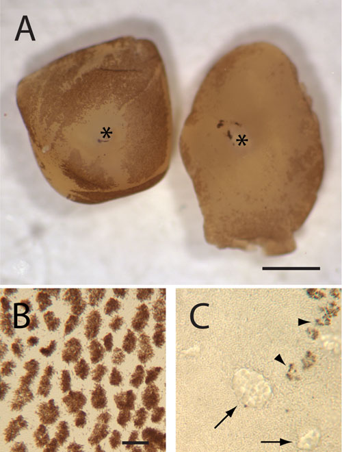
Figure 9. Neural retinas peeled from the eyes of 5 week-old p27+/- and p27-/- mice
A: Stereomicroscope view. Melanin pigment granules (dark brown) cover much of the outer surface of the hemizygous retina (left), except in the area surrounding the optic nerve (*). By contrast, the peeled nullizygous retina has relatively little adherent pigment in central regions, although some melanin is present in more peripheral regions (right). B and C: Bright-field micrographs. To obtain these images, the same retinas as seen in (A) were flat-mounted and typical regions viewed at high power. Melanin granule-containing microvillous processes (arrowheads in C), which have broken off during tissue separation, are present in much larger numbers in the hemizygous, as compared to the nullizygous, retina. Arrows indicate regions of outer nuclear layer dysplasia. A: Scale bar represents 1 μm. B and C: Scale bar represents 20 μm.