![]() Figure 7 of
Defoe, Mol Vis 2007;
13:273-286.
Figure 7 of
Defoe, Mol Vis 2007;
13:273-286.
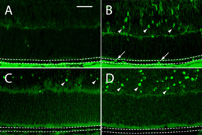
Figure 7. Cell proliferation and cell death in frozen sections of retinas from developing p27Kip1 hemizygous and nullizygous mice
A and C represent p27+/- and B and D represent p27-/- tissues. A and B: BrdU incorporation. Representative photomicrographs of the peripheral retina from animals injected on P7 and sacrificed on P8. Immunoreactive nuclei (arrows) are present in the nullizygous (B), but not hemizygous (A), RPE (between dotted lines). Note also the presence of BrdU-positive cells in the developing inner nuclear layer of the mutant retina (arrowheads). C and D: TUNEL assay. Apoptotic epithelial cells are undetectable in P8 retinal sections from both genotypes. On the other hand, TUNEL-positive cells are seen in the hemizygous neural retina and are especially plentiful in the nullizygous neural retina (arrowheads). Scale bar represents 40 μm.