![]() Figure 3 of
Defoe, Mol Vis 2007;
13:273-286.
Figure 3 of
Defoe, Mol Vis 2007;
13:273-286.
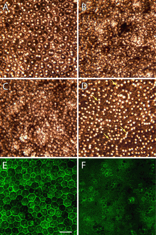
Figure 3. Flat-mounts of wild-type and mutant retinal pigment epithelium from P35 retinas
Whole-mounted retinal pigment epithelium (RPE) tissue from wild-type (A and E) and mutant (B, C, D, and F) P35 retinas were left unstained (A-F) or labeled with Alexa Fluor 488-phalloidin (E and F). In unstained preparations, the normal epithelium consists of relatively uniform polygonal cells with centrally-located nuclei (A). Mutant cells are, by contrast, smaller in diameter and less regular in their outline. While cellular density can vary somewhat, regions of the nullizygous RPE exhibiting a higher nuclear density also display increased cellular density (compare B, C, and D). In some mutant eyes, there are increased numbers of epithelial cells with nuclei displaced from central to peripheral regions (D, arrows). In 1 μm thick optical sections of Alexa Fluor 488-phalloidin-labeled tissues, apical cell boundaries are marked by rings of actin filaments associated with intercellular junctions, while microvillous processes are evident as punctate dots overlying the cytoplasm (E and F). Note the alignment and regularity of cell outlines in the wild-type RPE layer (E). The cytoskeleton of mutant epithelial cells, on the other hand, appears less well organized, and Alexa Fluor 488-phalloidin staining defines apical membrane compartments that are much smaller and more variable in diameter (F). Scale bar represents 40 μm.