![]() Figure 12 of
Defoe, Mol Vis 2007;
13:273-286.
Figure 12 of
Defoe, Mol Vis 2007;
13:273-286.
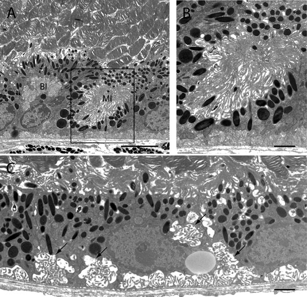
Figure 12. Electron micrographs of the RPE-photoreceptor interface from a p27Kip1-null retina
The low power image (A) shows an incompletely polarized epithelial cell displaying a microvillar inclusion (MI), as well as an inclusion of basolateral membrane (BI), within the cytoplasm of the cell. The boxed region in A is seen at higher power in B. The micrograph shown in C represents a region where there is an apparent expansion of the basal surface membrane, which also tends to occupy more central and lateral portions of cells (arrows). Such polarization defects occur in well-oriented regions of the retina. A: Scale bar represents 5 μm. B: Scale bar represents 1 μm. C: Scale bar represents 2 μm.