![]() Figure 1 of
Defoe, Mol Vis 2007;
13:273-286.
Figure 1 of
Defoe, Mol Vis 2007;
13:273-286.
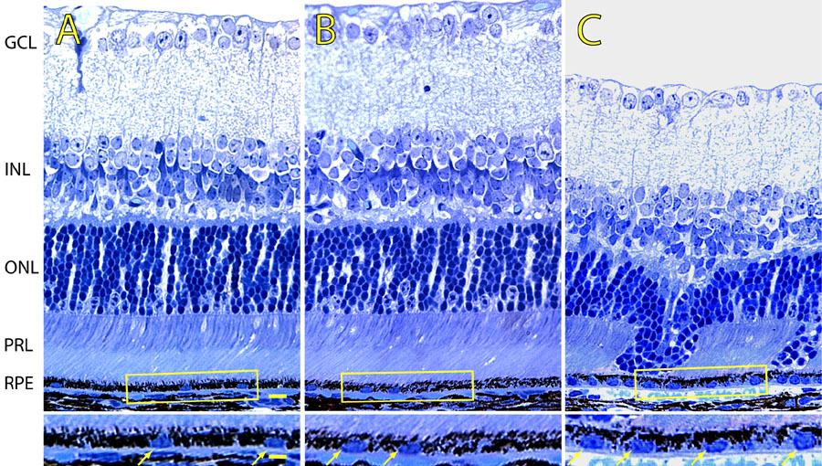
Figure 1. Light micrographs of JB-4 retinal sections derived from 5 week-old wild-type and p27Kip1 hemizygous and nullizygous mice
A represents p27+/+, B represents p27+/-, and C represents p27-/- retinal tissue. The RPE cell layer from p27Kip1-null animals appears thicker, with increased numbers of nuclei (arrows in insets). Also, in the mutant retina melanin granules tend to occupy more apical portions of the cytoplasm. Note that the dysplasia of the outer nuclear and photoreceptor layers is evident in the p27Kip1-null retina only. RPE represents retinal pigment epithelium; PRL represents photoreceptor layer; ONL represents outer nuclear layer; INL represents inner nuclear layer; GCL represents ganglion cell layer. Scale bar represents 10 μm. Inset scale bar represents 20 μm.