![]() Figure 6 of
McMahon, Mol Vis 2007;
13:258-272.
Figure 6 of
McMahon, Mol Vis 2007;
13:258-272.
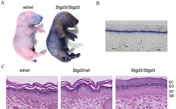
Figure 6. Defective skin barrier in neonatal homozygous Stgd3 mice
A: Skin permeability assay. Euthanized neonatal pups were immersed in a toluidine blue solution. The skin of homozygous Stgd3 mice, in contrast to their wt littermates, stained blue indicating a defect in skin barrier function. B: Light microscopy examination of a cross section of the dye-stained skin of the homozygous Stgd3 neonate shows penetration of the dye through the outer skin layers. C: Hematoxylin and eosin staining of homozygous Stgd3 skin shows the presence of all four epidermal layers: stratum corneum (SC), stratum granulosum (SG), stratum spinosum (SP) and stratum basale (SB). Note the more compact structure of stratum corneum in the mutant homozygous skin compared to the wt/wt and Stgd3/wt skin. Scale bar represents 100 μm in B.