![]() Figure 5 of
McMahon, Mol Vis 2007;
13:258-272.
Figure 5 of
McMahon, Mol Vis 2007;
13:258-272.
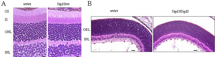
Figure 5. Light micrographs of hematoxylin and eosin stained sections of Stgd3 mouse retinas
A: Retinal sections from 8-month-old heterozygous Stgd3 mice and their wt littermates show similar thickness of photoreceptor outer segments (OS), inner segment (IS), outer nuclear (ONL) and inner nuclear (INL) layers. No significant changes in numbers and morphology of photoreceptors are evident. Scale bar represents 10 μm. B: Retinal sections from neonatal homozygous Stgd3 mice and their wt littermates show similar retinal histology, with distinct INL and larger outer retinal layers (ORL). The ORL contains the photoreceptor cell bodies which at this stage of development have not elaborated outer segments. Scale bar represents 100 μm.