![]() Figure 7 of
Reza, Mol Vis 2007;
13:18-30.
Figure 7 of
Reza, Mol Vis 2007;
13:18-30.
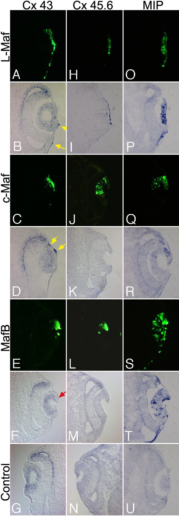
Figure 7. Maf misexpression stimulates Cx43, Cx45.6, and MIP in electroporated cells
Cryosections prepared from stage 16 embryos electroporated with wild-type L-Maf, c-Maf, and MafB were subjected to in situ hybridization with Cx43 (A-G), Cx45.6 (H-N), and MIP (O-U). Both L-Maf and c-Maf induced the expression of Cx43 (B,D, respectively, yellow arrows), while MafB suppressed Cx43 expression in the electroporated cells (F, red arrow). Cx 45.6 was activated by L-Maf only (I). c-Maf and MafB did not induce Cx45.6 (K,M, respectively). L-Maf induced the highest expression of MIP (P), followed by MafB (T) and c-Maf (R). Nonelectroporated contralateral eyes revealed that Cx45.6 (N) and MIP (U) were not expressed at this stage, while regular expression of Cx43 (G) was detected. Green fluorescence (GFP) indicates transgene expression (A,H,O,C,J,Q,E, L,S). This figure is representative of at least three independent experiments.