![]() Figure 4 of
Reza, Mol Vis 2007;
13:18-30.
Figure 4 of
Reza, Mol Vis 2007;
13:18-30.
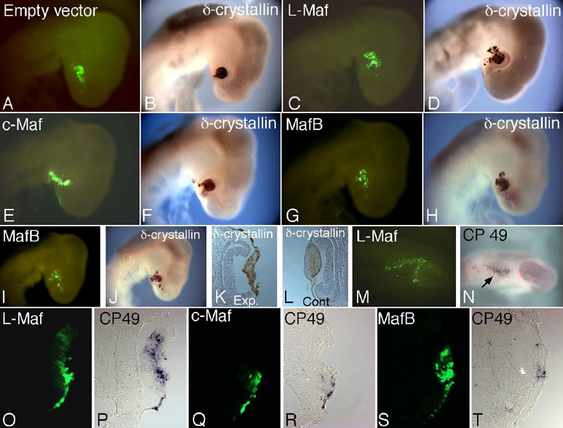
Figure 4. In vivo detection of δ-crystallin and CP49 by overexpressing L-Maf, MafB, and c-Maf, showing different inducing ability
Whole-mount immunostaining using δ-crystallin antibody was performed using stage 16 embryos electroporated with empty vector as negative control (A,B), wild-type L-Maf (C,D), c-Maf (E,F), and MafB (G-L) at stage 10. Embryo electroporated with empty vector showed no expression of δ-crystallin outside lens (B). Ectopic δ-crystallin expression was detected in all Maf-expressing embryos (D,F,H,J). However, it is apparent that L-Maf did induce δ-crystallin maximally (D). In most cases, MafB overexpression hampered lens shape (J,K). A section of the embryo shown in J revealed a lack of lens invagination and proper arrangement of cells (K). Nonelectroporated contralateral eye section showed normal expression of δ-crystallin and proper lens development (L). In similar experiments, cryosections from the eclectroporated embryos were subjected to in situ hybridization using Dig-labeled CP49 probe (O-T). L-Maf (O,P) strongly induced CP49, while c-Maf (Q,R) and MafB (S,T) showed lower activation of CP49. Further, whole-mount in situ hybridization was performed using L-Maf electroporated embryos after 5 h of incubation (M,N; stage 11), which revealed ectopic CP49 expression (N; arrow). Green fluorescence of GFP indicates transgene expression (A,C,E,G,I,M,O,Q,S). The figure is representative of at least three independent experiments. For whole-mounts, five to six embryos were used each time.