![]() Figure 3 of
Bentley, Mol Vis 2007;
13:237-242.
Figure 3 of
Bentley, Mol Vis 2007;
13:237-242.
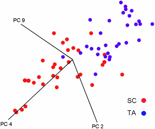
Figure 3. Three dimensional cluster plot of the spectral PCs for SC and TA cell populations
This figure shows a three dimensional cluster plot of the spectral PCs for SCs (red) and TA cells (blue). Nearness of points implies pattern recognition and the separation of sample clusters in the plots signifies structurally dissimilar groups. It is evident that the two populations of cells form distinct clusters, although the discrimination is not perfect.