![]() Figure 2 of
Bentley, Mol Vis 2007;
13:237-242.
Figure 2 of
Bentley, Mol Vis 2007;
13:237-242.
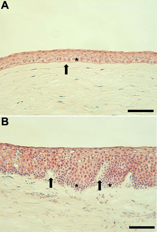
Figure 2. Light micrographs of the corneal and limbal epithelium
A: Light micrograph of the corneal epithelium. The TA cells are the basal cell layer (asterisk) below which is Bowman's layer (arrow). B: Light micrograph of the limbal epithelium. The invaginations of the basal lamina in the limbal region are termed the palisades of Vogt (arrow). The stem cells are located in the basal cell layer and are concentrated at the base of the rete ridge-like structures (asterisk). The scale bars are equal to 100 μm.