![]() Figure 3 of
Cheng, Mol Vis 2007;
13:2344-2352.
Figure 3 of
Cheng, Mol Vis 2007;
13:2344-2352.
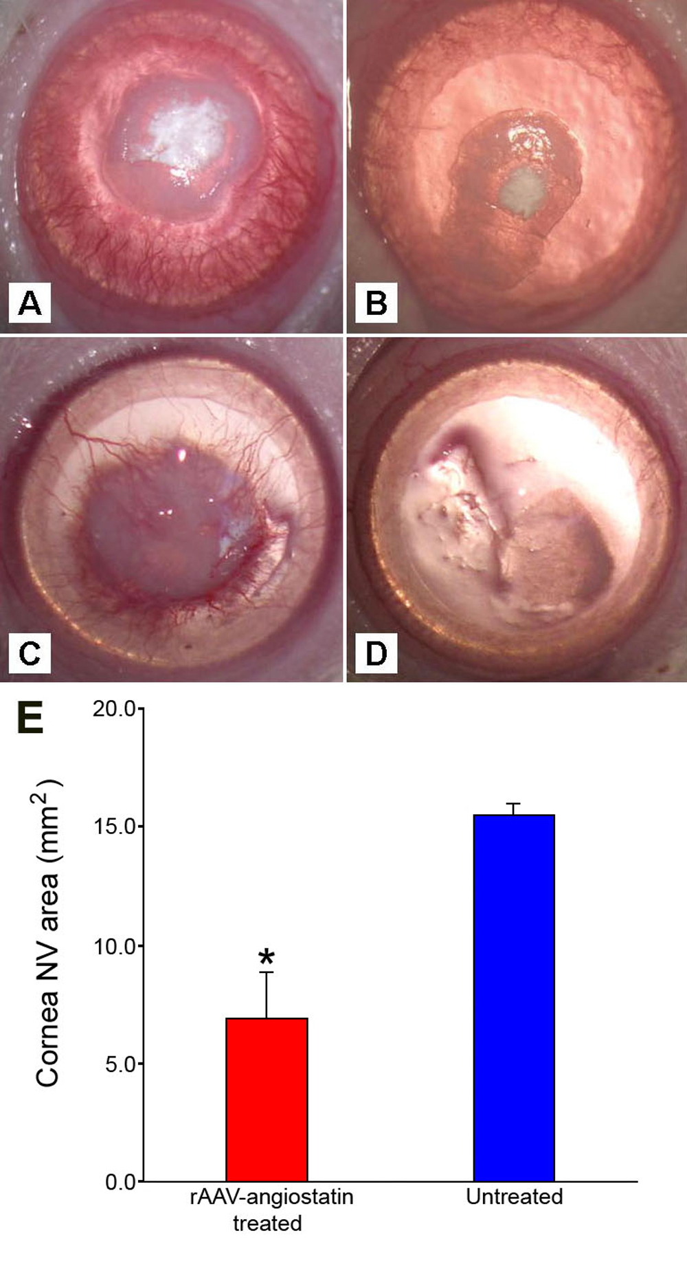
Figure 3. Effect of subconjunctival injection of rAAV-angiostatin on alkali burn-induced cornea neovascularization regression
One week before infliction of the alkali burn with 1 N NaOH in rats, the rats were assigned randomly either to a treatment group or control group. The treatment group received subconjunctival injection of rAAV-angiostatin three weeks before the alkali burn, and the control group received blank rAAV viral vector treatment. Corneal NV was examined daily by slit lamp for two weeks. Representative photographs of alkali burn-induced corneal NV at days 7 and 14 are shown. A: Dense NV is growing toward the central cornea. Seven days after the alkali burn, there was severe corneal opacity and epithelial defects in the rAAV viral vector control eye. B: The rAAV-angiostatin injected eye seven days after the alkali burn is shown. Note the scarcity of corneal NV and the mild to moderate corneal opacity. C: The rAAV viral vector control eye at day 14 follow-up is shown. Corneal NV and pannus maturation were evident. D: The rAAV-angiostatin injected eye 14 days after the alkali burn is shown. Not only the corneal NV was absent, but also the central corneal opacity was mild. E: The corneal NV area (mean±SD, n=6) measured at seven days after exposure to the alkali burn is shown. The rAAV-angiostatin treated group demonstrated significantly smaller NV area compared with the rAAV viral vector control group (the asterisk means that p<0.05).