![]() Figure 2 of
Cheng, Mol Vis 2007;
13:2344-2352.
Figure 2 of
Cheng, Mol Vis 2007;
13:2344-2352.
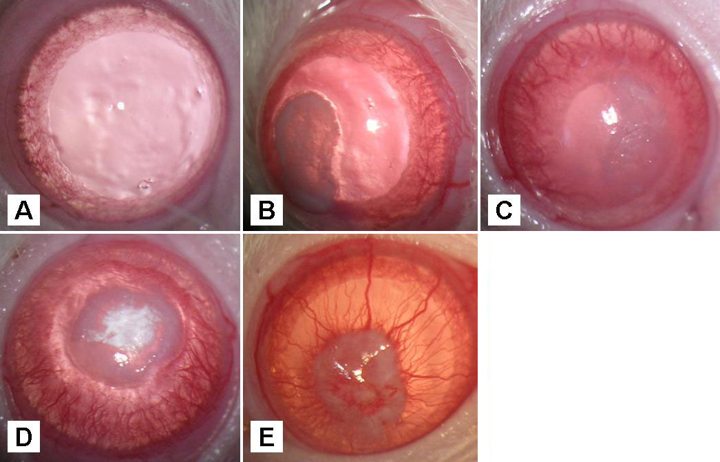
Figure 2. Alkali burn-induced corneal neovascularization in the rat animal model
Rats received alkali burns on the central cornea of their left eyes. Corneal NV was monitored by slit lamp examinations. A: In the Control treatment a normal and clear cornea was shown before induction of alkali burn. B:An opaque central cornea was observed after exposure to 1 N NaOH at PD0. C: At PD3, an initial peripheral NV ingrowth toward the central cornea with increased central corneal opacification was observed. D: At PD7, dense NV ingrowth, reaching the central cornea was observed. Note an elevated pannus surrounding the area of central epithelial defect. E: At PD14, cornea NV maturation with decreased vessels caliber was observed. Note that the central corneal scarring and pannus formation were stationary.