![]() Figure 1 of
Cheng, Mol Vis 2007;
13:2344-2352.
Figure 1 of
Cheng, Mol Vis 2007;
13:2344-2352.
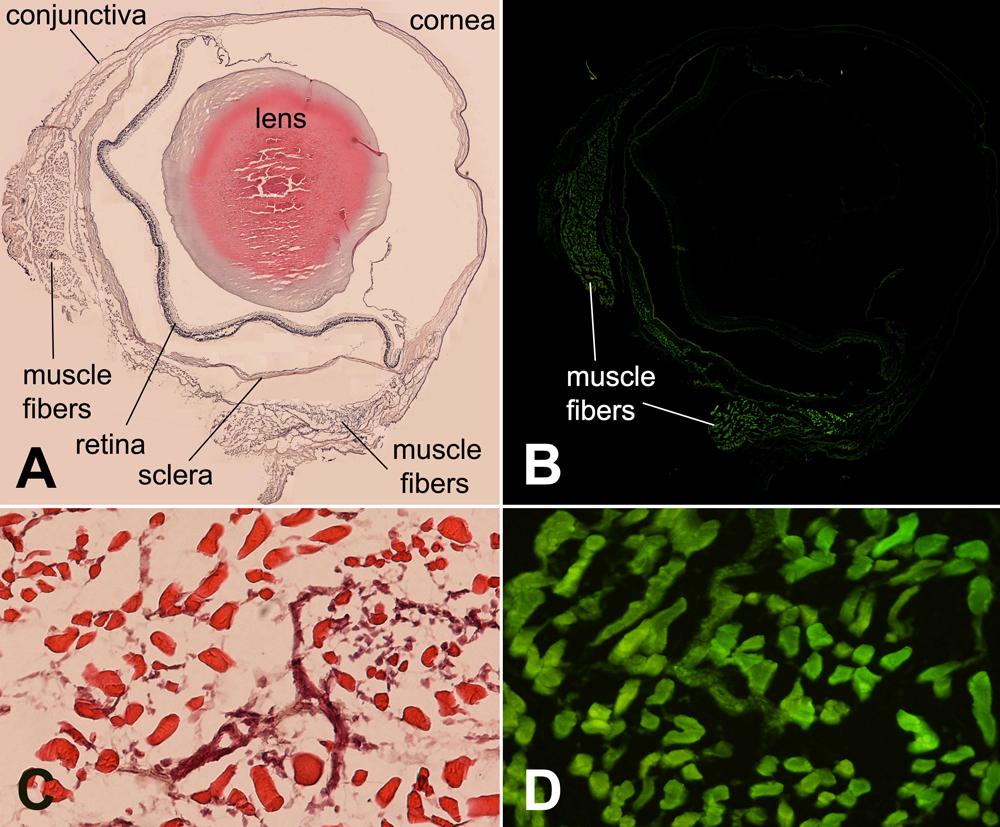
Figure 1. Histopathology and distribution of green fluorescent protein expression after subconjunctival injection of rAAV-GFP
Hematoxylin-eosin staining (A) and its corresponding fluorescent microscopy findings (B) of GFP overview expression in a rat eye three weeks after subconjunctival injection of rAAV-GFP is shown. GFP expression was distinctly detected in the muscle fibers adjacent to the EOM insertions. No evident green fluorescence was detectable in the conjunctiva, subconjunctival soft tissue, cornea, retina, choroid, or vitreous. Magnification, 40x. C: Hematoxylin-eosin staining of muscle fibers adjascent to EOM insertions is shown. Scarce inflammatory cell infiltration can be noted between the muscle fibers. D: Corresponding fluorescent microscopy findings of cryo-section in C. Note intense expression of GFP by the muscle fibers. Magnification, 400x.