![]() Figure 1 of
Young, Mol Vis 2007;
13:2328-2333.
Figure 1 of
Young, Mol Vis 2007;
13:2328-2333.
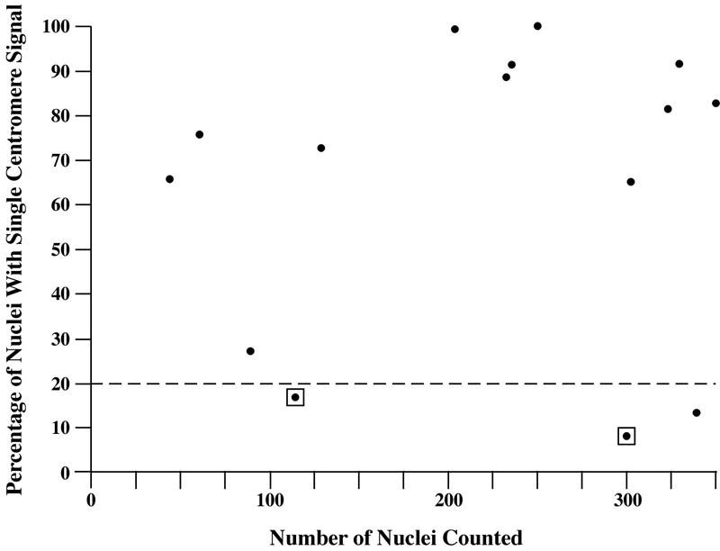
Figure 1. Monosomy 3 ratios by fluorescence in-situ hybridization in fine needle aspiration specimens
The scatter plot shows the distribution of scoring for the 15 samples determined to be monosomy 3 by fluorescence in-situ hybridization (FISH). The vertical axis represents sample heterogeneity with respect to monosomy 3. The 20% level is the threshold below which FISH and single nucleotide polymorphism (SNP) mapping array results diverge. Boxed data points indicate monosomy 3 by FISH which is inconsistent with mapping array.