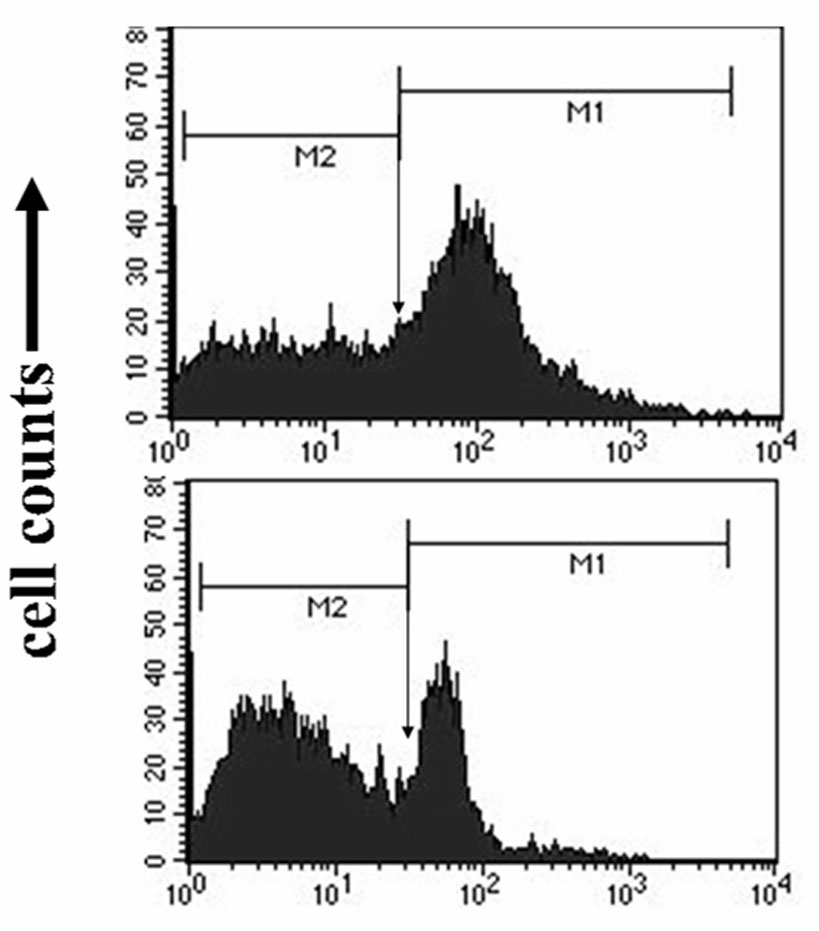
Figure 3. Representative flow cytometry data Counts to right of arrow represent population of ARPE-19 containing fluorescein isothiocyanate-labeled
rod-outer- segments. The area under the curve to the right of the arrow is greater in retinal pigment epithelium cultured
on younger Bruch’s membrane (top panel, from a 14-year-old donor) than on older Bruch’s membrane (bottom panel, from a 76-year-old
donor).

 Figure 3 of
Sun, Mol Vis 2007; 13:2310-2319.
  Figure 3 of
Sun, Mol Vis 2007; 13:2310-2319.