Figure 2. Binding and uptake of fluorescein isothiocyanate -rod outer segment particles by ARPE-19 are decreased when cultured on older
Bruch’s membrane A: Phase contrast microscopic image shows ARPE-19 two- weeks after seeding onto a 16-year-old donor Bruch’s membrane. B: Phase contrast image shows ARPE-19 two weeks after seeding onto an 81-year-old donor Bruch’s membrane. C: Fluorescence image of ARPE-19 on a 16-year- old donor Bruch’s membrane fed with labeled rod-outer-segments shows cells border
(broken arrows) and intense labeling of ingested outer segments (solid arrow). D: Fluorescence image of ARPE-19 on an 81-year-old donor Bruch’s membrane fed with labeled rod-outer-segments shows cells border
(broken arrows) and less labeling of ingested outer segments (solid arrow) (compare C to D).
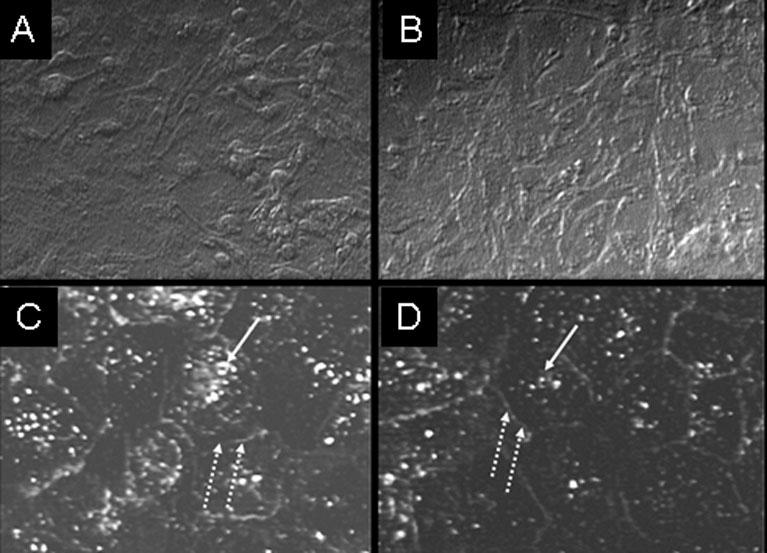
 Figure 2 of
Sun, Mol Vis 2007; 13:2310-2319.
Figure 2 of
Sun, Mol Vis 2007; 13:2310-2319.