Figure 1. ZO-1 immune staining of ARPE-19 cells cultured to confluence on Bruch’s membrane Fluorescent microscopy shows ZO-1 immune
staining of ARPE-19 cells cultured for two weeks until they reached confluence on (A) a 20-year-old Bruch’s membrane and (B) an 80-year-old Bruch’s membrane. Note the hexagonal shape of the ARPE-19 monolayer, with no apparent morphological difference
between ARPE-19 cultured on younger versus older Bruch’s membrane.
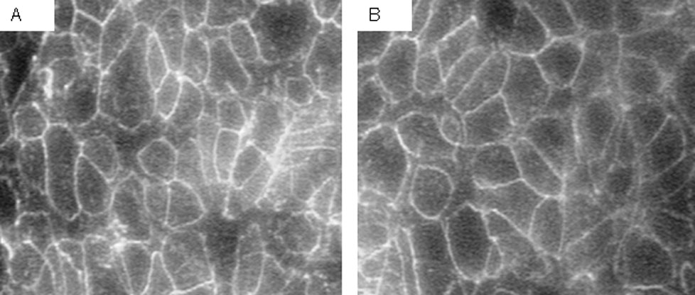
 Figure 1 of
Sun, Mol Vis 2007; 13:2310-2319.
Figure 1 of
Sun, Mol Vis 2007; 13:2310-2319.