![]() Figure 2 of
Murugesan, Mol Vis 2007;
13:2301-2309.
Figure 2 of
Murugesan, Mol Vis 2007;
13:2301-2309.
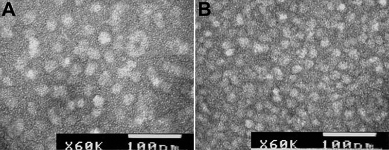
Figure 2. Electron microscopic image of αAG98R and wild-type αA-crystallin
A drop of 1 mg/ml protein was negatively stained with uranyl acetate and examined under JEOL 1200EX electron microscope. The bar in the micrograph is 100 nm. A: α?AG98R; B: wild-type αA-crystallin.