![]() Figure 4 of
Zhang, Mol Vis 2007;
13:2289-2300.
Figure 4 of
Zhang, Mol Vis 2007;
13:2289-2300.
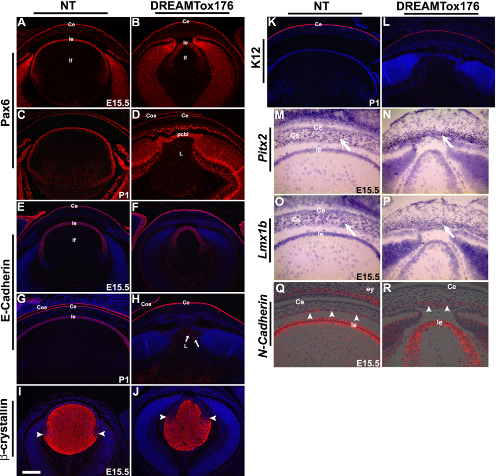
Figure 4. Expression of corneal and lens markers in DREAM-Tox176 (OVE1757) mice.
Immunohistochemistry was performed on E15.5 or P1 ocular sections to detect expression of Pax6 (A-D), E-cadherin (E-H), β-crystallin (I,J), and Keratin-12 (K12; K,L). Antigen-antibody complexes are in red and DAPI staining (blue) reveals the nuclei. DAPI staining is not shown in A-D. In situ hybridizations were performed on sections of E15.5 or P1 heads from nontransgenic (NT), and DREAM-Tox176 (OVE1757) mice using [35S]-labeled riboprobes for Pitx2 (M,N), Lmx1b (O,P), and N-Cadherin (Q,R). Only bright-field images are shown in panels M-P and silver grains (arrows) appear black. In Q and R, silver grains were pseudo-colored red and overlaid on top of the respective bright-field images. Pitx2 and Lmx1b, genes normally expressed in the ocular mesenchymal cells showed elevated expression in the layer of mesenchymal cells directly adjacent to the lens epithelium (N and P). Expression of N-Cadherin, a marker for differentiated corneal endothelial cells, could be seen at E15.5 (R). The staining in the core of the lens in M and O (asterisk) is an artifact of the dark-field illumination. Abbreviations: Ce, corneal epithelium; Coe, conjunctival epithelium; Cs, corneal stroma; L, lens; le, lens epithelial cells; lf, lens fiber cells; NT, nontransgenic mice; pcbi, presumptive ciliary body and iris. The scale bar (I) represents 60 μm in A-L and 30 μm in M-R. The scale bar (O) represents 100 μm in K-P.