![]() Figure 1 of
Li, Mol Vis 2007;
13:2282-2288.
Figure 1 of
Li, Mol Vis 2007;
13:2282-2288.
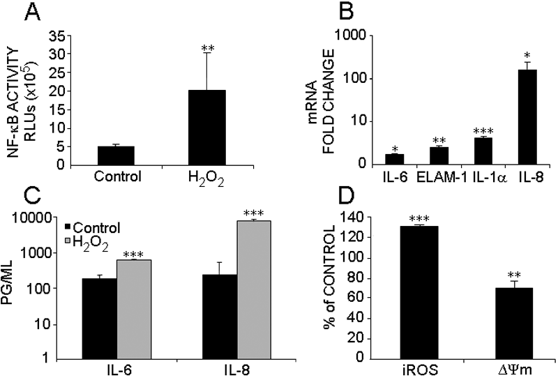
Figure 1. Induction of proinflammatory factors by chronic H2O2 treatment
TM cells were treated with H2O2 200 μM twice a day for four days and then they were allowed three days to recover. A: Activation of NF-κB measured by NoShift II NF-κB assay. Data represents the mean values of NF-κB activation in relative light units (rlu) ±SD of n=3. B: Realtime Q-PCR analysis of the induction of IL-1α, IL-6, IL-8, and ELAM-1. For each individual sample, the expression level of each gene was first normalized with that of β-actin and then the relative differences between groups were expressed as mean fold changes compared with the controls ±SD of n=3. C: Protein levels of IL-6 and IL-8 in cell culture media assessed by ELISA. Data represent means of protein concentration ±SD of n=6. D: Induction of iROS and decrease of Δψm detected by H2DCFDA and JC-1. Data showed a percentage of control ±SD (n=5) in iROS treated with H2O2. This increase in iROS was associated with a reduction of percentage ±SD in Δψm (n=3). An asterisk means that p<0.05; a double asterisk means that p<0.01; and three asterisks mean that p<0.001 (compared to nontreated control).