Figure 4. An X-ray diffraction pattern from a Xenopus interphotoreceptor retinol-binding protein crystal recorded at the A-1 station of the Cornell High Energy Synchrotron Source.
The detector was at a distance 250 mm; the wavelength, the oscillation angle and the exposure time were 0.978 Å, 1°, and 15
s, respectively. The right upper corner segment is shown in higher contrast to demonstrate the limiting resolution of diffraction
of ~2.45 Å.
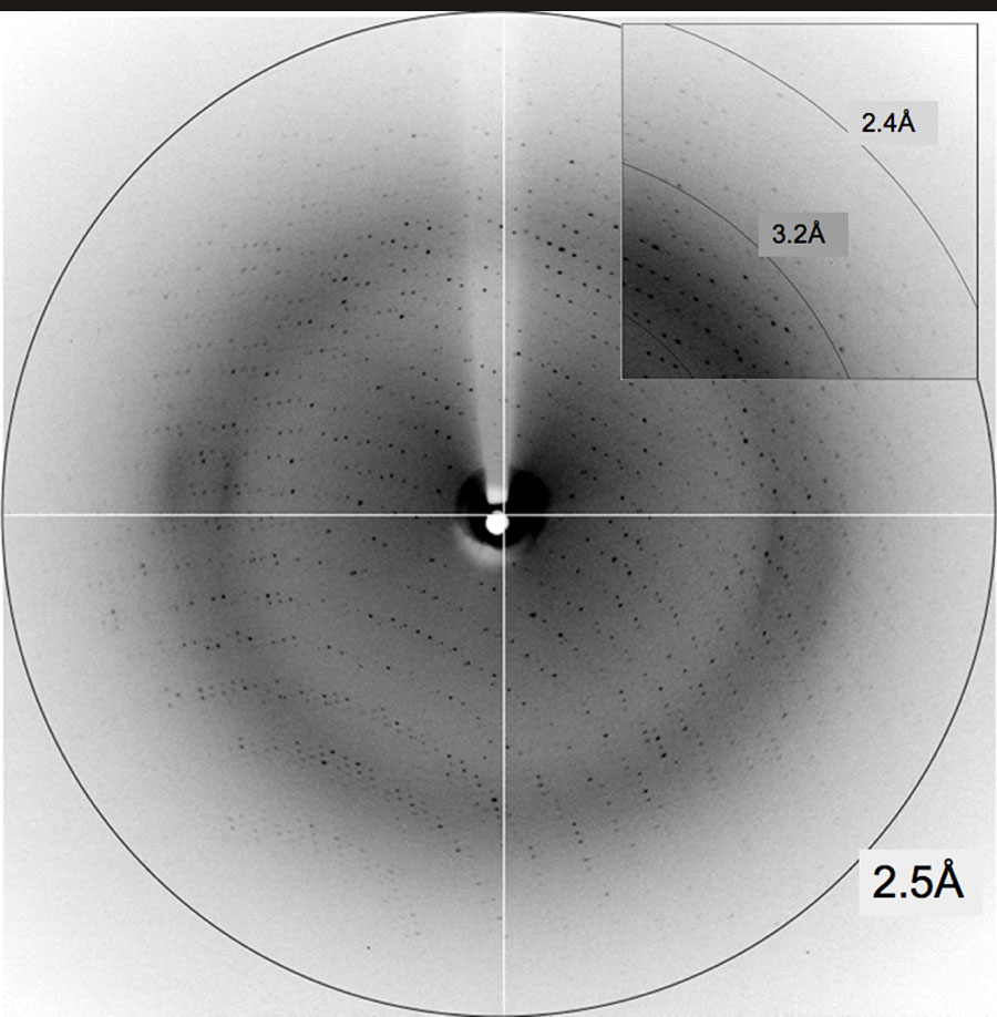
 Figure 4 of
Ghosh, Mol Vis 2007; 13:2275-2281.
Figure 4 of
Ghosh, Mol Vis 2007; 13:2275-2281.