![]() Figure 7 of
Camelo, Mol Vis 2007;
13:2263-2274.
Figure 7 of
Camelo, Mol Vis 2007;
13:2263-2274.
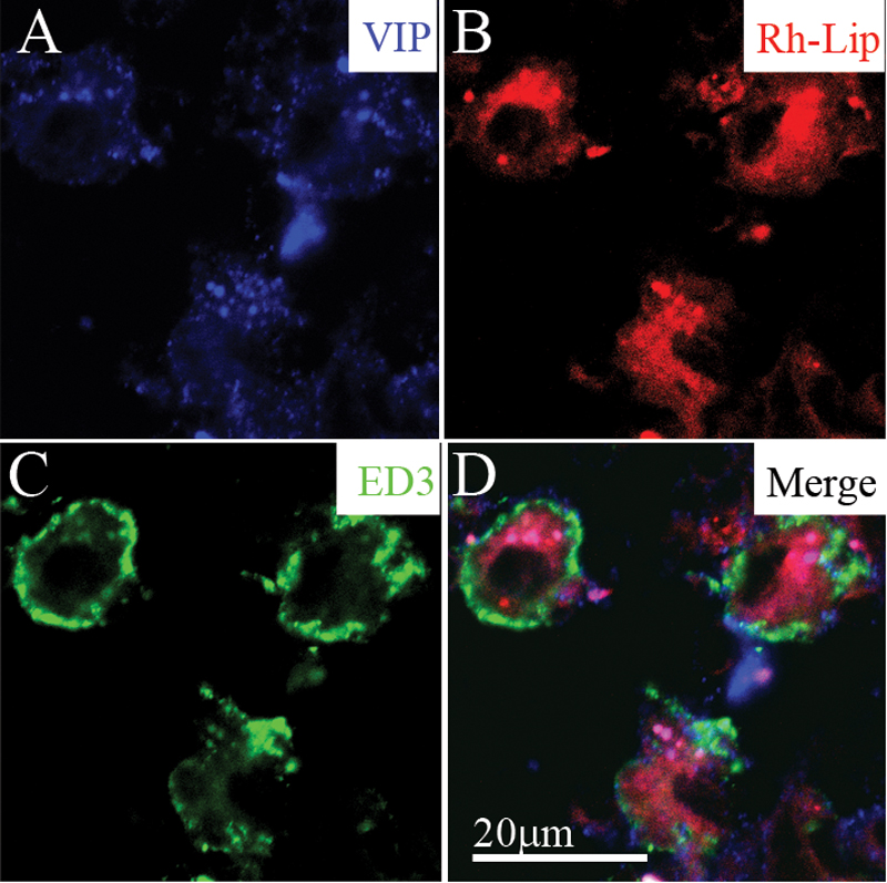
Figure 7. VIP-loaded rhodamine-conjugated-liposomes (VIP-Rh-lip) internalization and VIP expression by ED3-positive macrophages in cervical LN 24 h following IVT injection of VIP-Rh-Lip
A: Free VIP, detected with rabbit anti-VIP antibody (blue) localized within cells containing Rh-Lip (red; B) and expressing ED3 green (C). D: Merge image showing membranous expression of ED3 by cells containing Rh-lip and blue granules within liposomes. The bar in D represents 20 μm in all images. Confocal microscopy optical section is 2 μm in all images. Representative images of two experiments performed on cervical LN from two rats.