![]() Figure 6 of
Camelo, Mol Vis 2007;
13:2263-2274.
Figure 6 of
Camelo, Mol Vis 2007;
13:2263-2274.
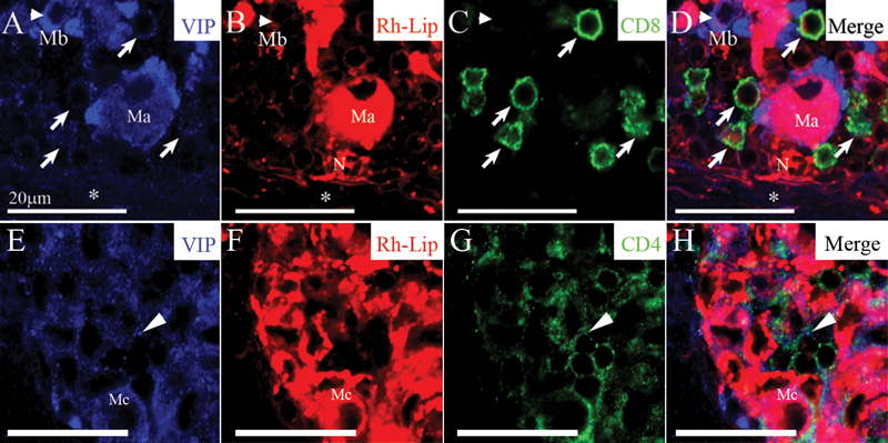
Figure 6. VIP detection in CD4 and CD8-positive T-cells in the cervical LN of rats receiving an IVT injection of VIP-loaded-rhodamine-conjugated-liposomes
A-D: Immuno-detection of VIP as blue dots in the LN capsule (asterisk), the LN parenchyma, within VIP-Rh-Lip-bearing macrophage (Ma) but also within macrophages that do not contain VIP-Rh-Lip (Mb, arrowhead): compare the localization of VIP in A, with the localization of VIP-Rh-Lip in red (B). VIP is present at the membrane of CD8-positive T lymphocytes in green (arrows): compare image A and C. Note the presence of a neutrophil (N) in B and D containing Rh-Lip in contact with macrophage Ma. VIP is present in small cells expressing CD4 in green (arrows; G,H) in the vicinity of macrophages containing Rh-Lip (Mc; E,F). In A to H, VIP is in blue, Rh-Lip in red, T cell markers in green and colocalization is in purple. All bars represent 20 μm, confocal microscopy optical section is 1.5 μm in all images. Representative images of two experiments performed on cervical LN from two rats.