![]() Figure 5 of
Camelo, Mol Vis 2007;
13:2263-2274.
Figure 5 of
Camelo, Mol Vis 2007;
13:2263-2274.
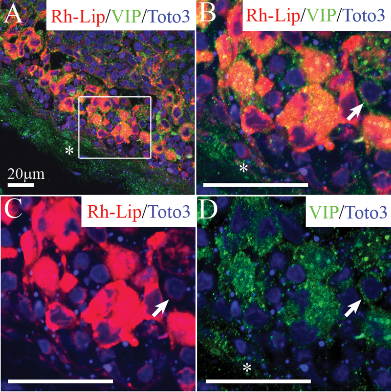
Figure 5. VIP biodistribution in cervical LN 24 h following IVT injection of VIP-Rh-Lip
A: Free VIP (green), detected with rabbit anti-VIP antibody is localized in the sinus (asterisks) and within subcapsular sinus macrophages containing rhodamine-conjugated-liposomes (red; B to D) Enlargement of the inset in A is shown in B to D. Free soluble VIP (green) is detected as green dots in the LN capsule (asterisks) and the LN parenchyma. VIP (appearing as yellow granules) is also detected within resident macrophages containing Rh-Lip (red cells in B and C). VIP is detected in small round cells that contain only minute amounts of Rh-Lip (arrows in B, C, and D). In A to D VIP appears green, Rh-Lip appears red and colocalization is indicated in yellow. Nuclei are stained in blue with Toto3®-iodide.All bars represent 20 μm, confocal microscopy optical section is 1.5 μm in all images. Representative images of two experiments performed on cervical LN from two rats.