![]() Figure 3 of
Camelo, Mol Vis 2007;
13:2263-2274.
Figure 3 of
Camelo, Mol Vis 2007;
13:2263-2274.
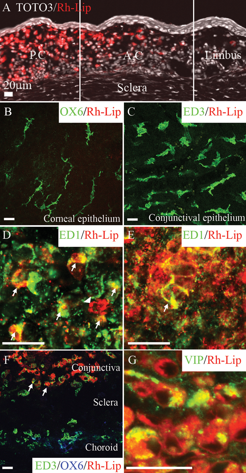
Figure 3. rhodamine-conjugated-liposomes and VIP biodistribution in conjunctiva 24 h following IVT injection of Rh-Lip and VIP-Rh-Lip
A: Following IVT injection, Rh-Lip (red) are internalized by cells in the conjunctival stroma. Fluorescent liposomes are not detected in conjunctival epithelium and sclera. Liposome concentration is maximal in the posterior conjunctiva (P.C) near the site of injection and decreases toward the anterior conjuntiva (A.C.) and the limbus. No liposomes were detected in the corneal stroma (data not shown). Nuclei stained with Toto3®-iodide are depicted in white. B: OX6-positive dendritic cell in the epithelium and C: ED3-positive macrophages in the conjunctival epithelium do not internalize Rh-Lip. D: ED1-positive cells in the anterior conjunctival stroma and E: in the posterior conjunctival stroma contain large amount of Rh-Lip. F: Fluorescent Rh-Lip (red) in the conjunctival stroma are taken up by ED3-positive (green), OX6 (blue)-negative macrophages (arrows). Choroidal ED3 and OX6-positive cells do not contain liposomes. G: VIP expression in the conjunctiva of a rat that received an IVT injection of VIP-Rh-Lip. VIP, detected with rabbit anti-VIP antibody (green) is localized in cells containing Rh-Lip (red) in the conjunctiva. Note the extra cellular green dots representing free VIP not internalized by cells. VIP appears green, Rh-Lip appears red and colocalization is indicated in yellow. All bars represent 20 μm, confocal microscopy optical section is 1.5 μm in all images. Photographs are representative of images obtained from frozen sections (A,F,G) and whole mounts (B-E) performed on a total of 12 eyes except G that is a representative image of ocular frozen sections from four eyes.