![]() Figure 2 of
Camelo, Mol Vis 2007;
13:2263-2274.
Figure 2 of
Camelo, Mol Vis 2007;
13:2263-2274.
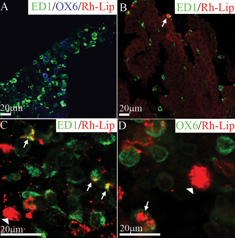
Figure 2. Biodistribution and phenotype of cells internalizing rhodamine-conjugated-liposomes in the iris and ciliary body of normal rats 24 h post IVT injection of Rh-Lip
A: On a frozen section of the iris, resident iridal ED1-positive (green) and OX6-positive cells (blue) are negative for fluorescent liposomes. B: Frozen section of the ciliary body showing a rare ED1-positive cell containing liposomes (arrow), ED1 green, rhodamine-conjugated-liposomes red, colocalization yellow. C: Ciliary body flat mount with ED1-positive cells containing fluorescent liposomes (arrows). The white arrowhead shows liposomes internalized by an ED1-negative cell. Other fluorescent liposomes appear not to be internalized by cells (red dots). D: Uptake of rhodamine-conjugated-liposomes by OX6-positive cells (green) appears limited to a few cells (arrow), the arrowhead shows liposomes internalized inside an OX6-negative cell. Photographs are representative of images obtained from frozen sections (A,B) and whole mounts (C,D) performed on a total of 12 eyes. All bars represent 20 μm. Confocal optical section, in all images, is 1.5 μm.