![]() Figure 2 of
Banh, Mol Vis 2007;
13:2248-2262.
Figure 2 of
Banh, Mol Vis 2007;
13:2248-2262.
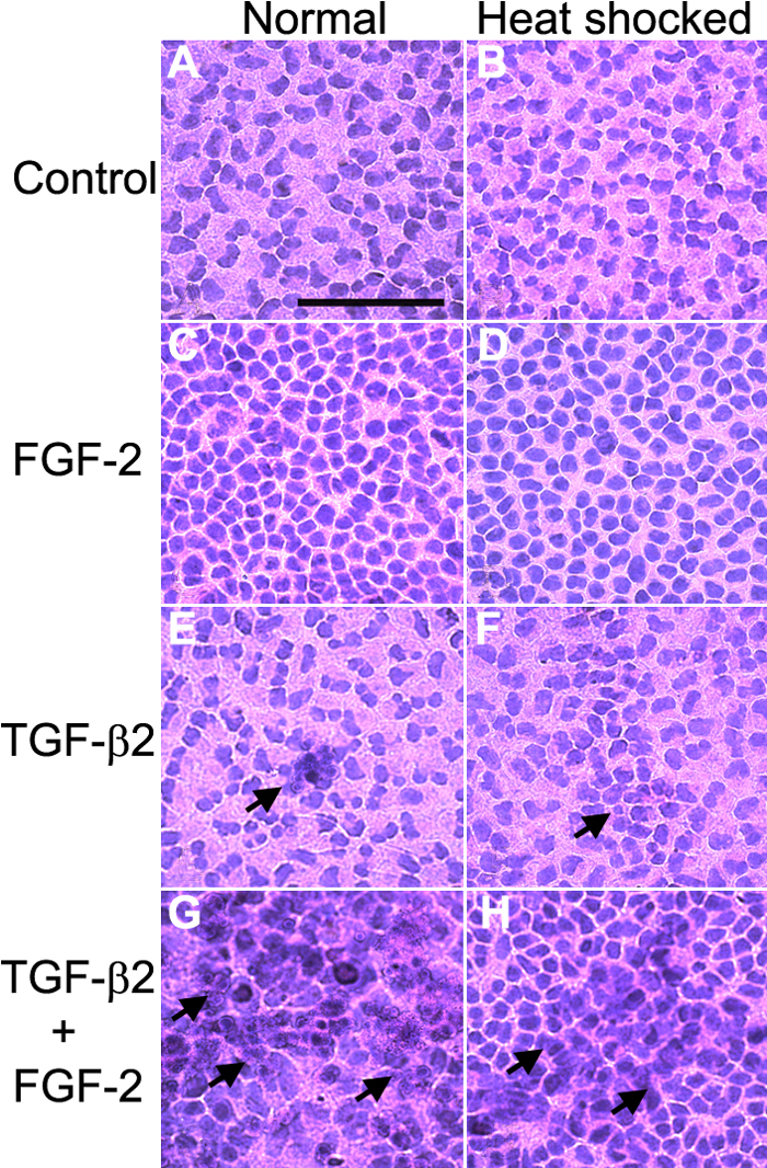
Figure 2. Histological analysis of rat lens epithelial explants
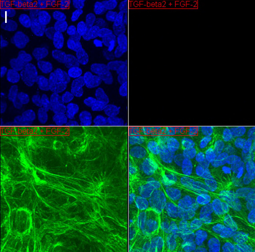
Control (A and B), FGF-2 (C and D), TGF-β2 (E and F), and TGF-β2/FGF-2 (G and H) lens epithelial explants under normal conditions (A, C, E, and G) and after heat shocked treatment (B, D, F, and H) are shown (n=4 for each treatment group). Control epithelial cells (A and B) show normal morphology. FGF-2 treated explants (C and D) have rounder cells and also show greater cell density than the control explants. Treatment with TGF-β2 (E-H) induced formation of multilayer plaques (arrows). The simultaneous treatment of epithelial explants with TGF-β2 and FGF-2 (G and H) induced the formation of considerably larger and more diffused plaques than TGF-β2 (E and F) alone. Both the heat shocked and normal cultured explants show similar phenotype. However, the plaques formed on the heat shocked TGF-β2/FGF-2 explants are less severe and less expansive than the non-heat shocked TGF-β2/FGF-2 plaques. The scale bar represents 100 μm. I: The video (below) demonstrate a confocal z-stack on a y-axis projection of a TGF-β2/FGF-2 treated explant stained for α-SMA (green) and nuclei (DAPI:blue). This video shows multiple cell layers with significant α-SMA staining.
Note that the slide bar at the bottom of the quicktime movie can be used to manually control the flow of the movie. If you are unable to view the movie, a representative frame is included below.
| This animation requires Quicktime 6 or later. Quicktime is available as a free download. |