![]() Figure 1 of
Banh, Mol Vis 2007;
13:2248-2262.
Figure 1 of
Banh, Mol Vis 2007;
13:2248-2262.
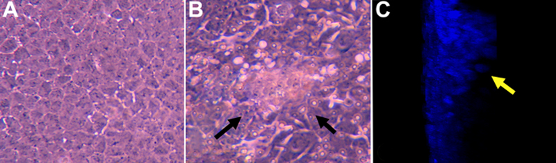
Figure 1. Cultured rat lens epithelial explants
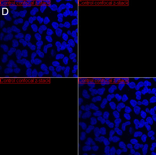
The phase contrast photographs represents a control (A) and a TGF-β2/FGF-2 treated lens epithelial explant (B). C shows a confocal z-stack section (a y-axis projection) of a TGF-β2/FGF-2 treated lens epithelial explant. The control rat explant demonstrates a regular monolayer of lens epithelial cells while the TGF-β2/FGF-2 treated explants (B and C) formed mutilayer plaques (arrows). D: The video (below) demonstrates a confocal z-stack on a y-axis projection of a control explant stained for α-SMA (green) and nuclei (DAPI:blue). This video shows a single layer of cells with no staining α-SMA.
Note that the slide bar at the bottom of the quicktime movie can be used to manually control the flow of the movie. If you are unable to view the movie, a representative frame is included below.
| This animation requires Quicktime 6 or later. Quicktime is available as a free download. |