![]() Figure 8 of
Park, Mol Vis 2007;
13:2222-2232.
Figure 8 of
Park, Mol Vis 2007;
13:2222-2232.
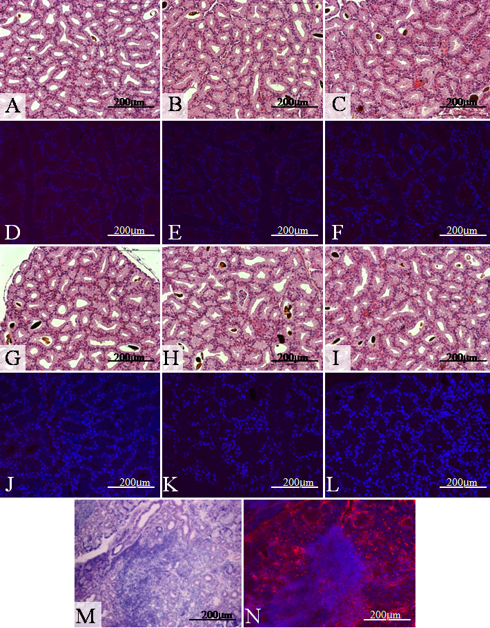
Figure 8. Histologic analysis
Hematoxylin and eosin (H&E) staining of lacrimal glands showed preservation of normal acinar structure and a lack of inflammatory cell infiltration in three specimens from two weeks (A-C) and another three specimens from four weeks (G-I). Immunofluorescent staining with CD3e monoclonal antibody showed no significant T lymphocyte infiltration in all specimens (D-F and J-L). H&E and immunofluorescent staining of lacrimal glands from MRL/Mpj mice as a positive control showed diffuse T lymphocytes infiltration (red) with nuclear counter staining (blue; M and N). A and D: control, B and E: saline injection, C and F: BTX-B injection, G and J: artificial tear treatment, H and K: cyclosporin A treatment, I and L: flurometholone treatment.