![]() Figure 2 of
Hanna, Mol Vis 2007;
13:2194-2208.
Figure 2 of
Hanna, Mol Vis 2007;
13:2194-2208.
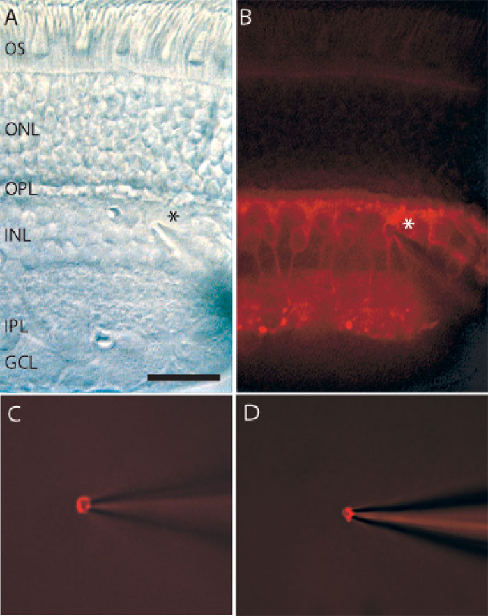
Figure 2. Labeling and extraction of rod bipolar cells
A: Nomarski-DIC image shows a 60-80 μm thick vertical section through macaque retina. A patch pipette electrode was positioned near the inner nuclear layer (INL), indicated by asterisk. B: The same retinal slice under fluorescence shows immuno-label of rod bipolar cells with antibodies against PKCα; their axons terminate just at the border of the inner plexiform layer (IPL) with the ganglion cell layer (GCL). C, D: Two representative single cells labeled for PKCα are shown following removal with patch pipette electrode. The retinal slice is no longer visible beyond the plane of focus. The following abbreviations are in effect: outer segments (OS), outer nuclear layer (ONL), outer plexiform layer (OPL). Scale bar equals 20 μm for both A and B.