![]() Figure 8 of
Mojumder, Mol Vis 2007;
13:2163-2182.
Figure 8 of
Mojumder, Mol Vis 2007;
13:2163-2182.
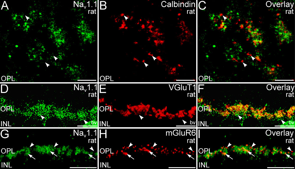
Figure 8. Analysis of single confocal optical sections indicated that Nav1.1 labeling is present in multiple locations in the outer plexiform layer
A-C: Confocal microscopy of double labeling for Nav1.1 and calbindin at the level of the OPL in retinal wholemount confirmed that little Nav1.1 labeling was present in the tips of horizontal cell dendrites (labeled for calbindin; arrowheads). A single optical section is shown. D-F: Labeling for Nav1.1 and VGluT1 co-stratifies in the OPL, suggesting some Nav1.1 labeling was associated with photoreceptor terminals. However, a substantial amount of Nav1.1 labeling did not colocalize with VGluT1 labeled photoreceptor terminals (arrowheads). Labeling of blood vessels (bv) was non-specific. A single confocal optical section shown. G-I: Only limited colocalization of Nav1.1 and mGluR6 labeling on the dendritic tips of ON bipolar cells is present in the OPL (arrows). Although most mGluR6-positive puncta did not show colocalization with Nav1.1 (arrowheads), labeling for Nav1.1 and mGluR6 were found in close proximity. A single confocal optical section is shown. The following abbreviations were used: outer nuclear layer (ONL); inner nuclear layer (INL). Scale bars equal 5 μm for A-C; 10 μm for D-I.