![]() Figure 6 of
Mojumder, Mol Vis 2007;
13:2163-2182.
Figure 6 of
Mojumder, Mol Vis 2007;
13:2163-2182.
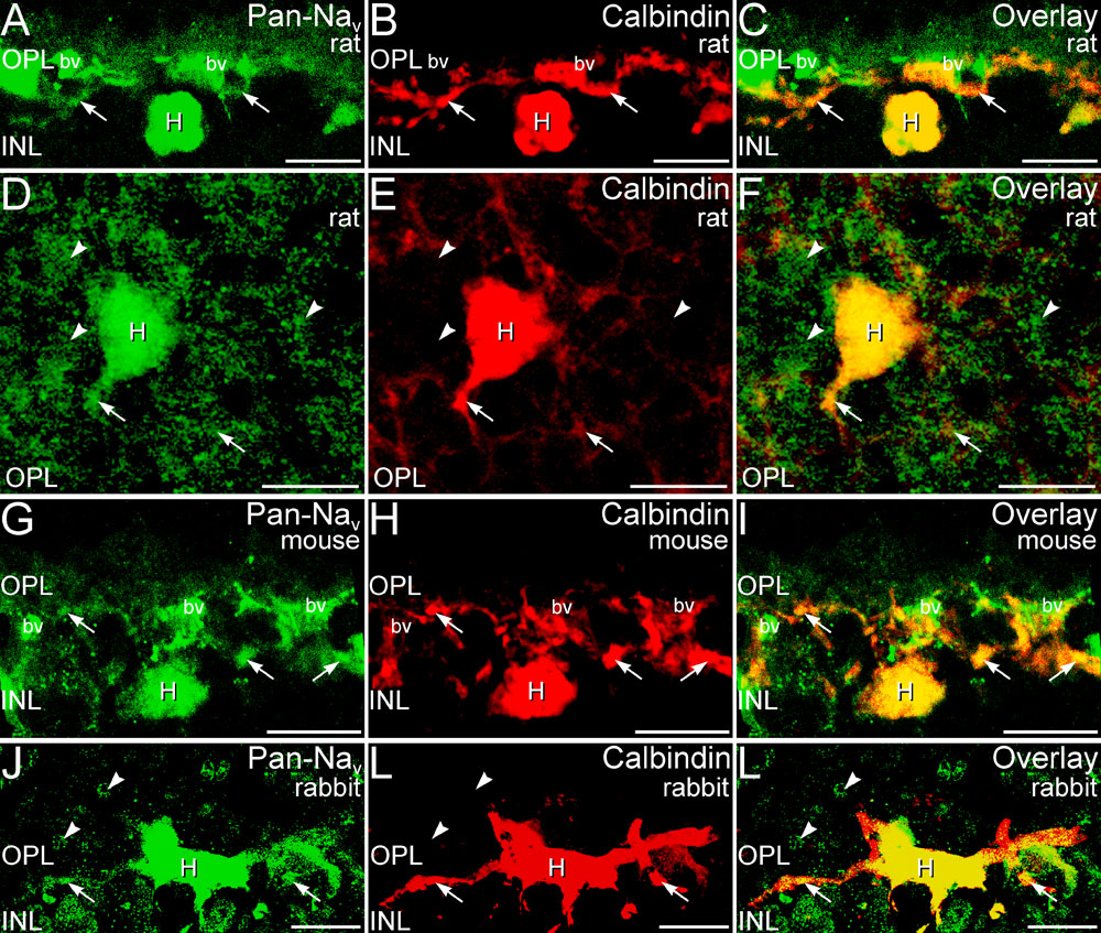
Figure 6. Horizontal cells showed Pan-Nav labeling
Double labeling for Pan-Nav (green) and the horizontal cell marker calbindin (red) confirmed that horizontal cells (H) and their processes (arrows) expressed Nav1 α-subunits in rat retina (A-C: vertical section; D-F: wholemount), mouse retina (G-I), and rabbit retina (J-L). Additional Pan-Nav labeling that is not associated with calbindin-positive horizontal cell processes also is present in the outer plexiform layer (OPL) and was particularly visible in whole mounted retina (arrowheads in D-F). Distinctive annuli of Pan-Nav labeling (arrowheads in J-L) were noted in the rabbit OPL. Labeling of blood vessels (bv) in mouse and rat retina was non-specific. In the figure inner nuclear layer is abbreviated INL. Scale bars equal 10 μm for A-F; 20 μm for G-L.