![]() Figure 5 of
Mojumder, Mol Vis 2007;
13:2163-2182.
Figure 5 of
Mojumder, Mol Vis 2007;
13:2163-2182.
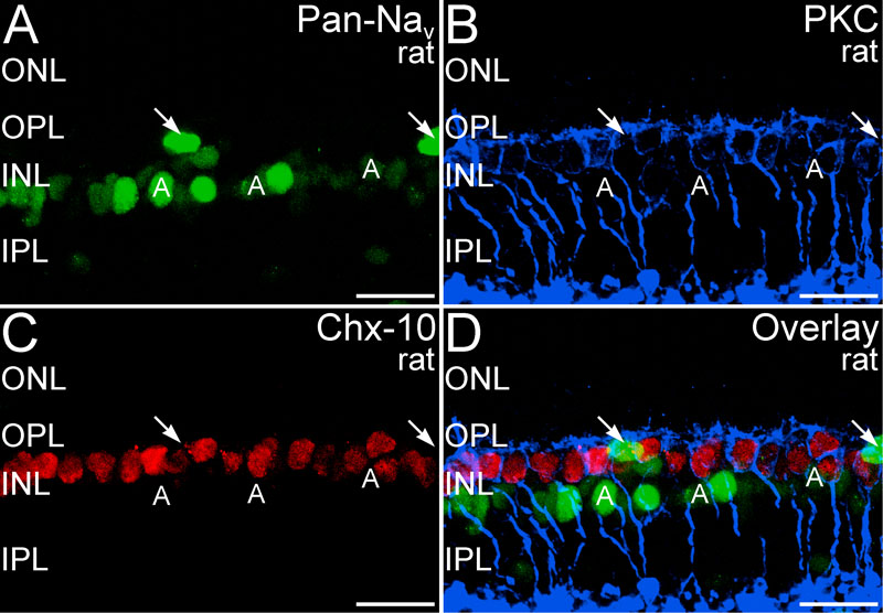
Figure 5. Pan-Nav labeling was absent from bipolar cell bodies
A-D: Triple labeling for Pan-Nav (green), the rod bipolar cell marker protein kinase C (PKC, blue) and the pan-bipolar cell marker Chx-10 (red), showed no colocalization of Pan-Nav labeling with bipolar cell markers. A: Pan-Nav labeling was present in many amacrine cell bodies (A) in the proximal inner nuclear layer (INL) and a few cell bodies in the distal INL (arrows). B: Rod bipolar cells showed labeling for PKC throughout the cell. C: Labeling for Chx-10 is present in the nuclei of all bipolar cells. Overlay of panels A-C showing that Pan-Nav labeling did not colocalize with labeling for either PKC or Chx-10. Images are a projection of 53 optical planes; 7.2 μm total thickness. Rat retina is shown. The following abbreviations were used: outer nuclear layer (ONL); inner plexiform layer (IPL). Scale bar equal 20 μm.